Networks of Neurons and Synapses
We continue from where we left off. Revisit the neuron structure.
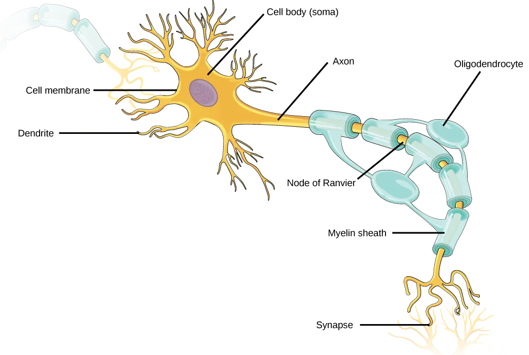 Courtesy: Openstax
With Neuron Doctrine coming out as best representation of our nervous system. We understood that each neuron function as discrete units with different forms and structure. It is estimated that a human have around 86 billion neurons in the human brain, with estimates ranging between 61 and 99 billion. Neurons are connected using synapse.
Synapse
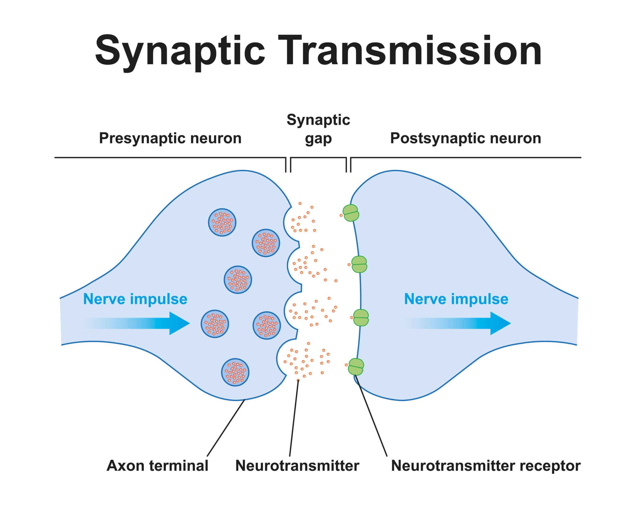 Source: ALI DAMOUH/SCIENCE PHOTO LIBRARY / Getty Images
Synapses is the connection point of the nervous system, allowing neurons to form networks. A single neuron isn't always a 1-1 connection, some are 1-n connection (where n > 1 > 100000). Neurons are connected to nearby neurons, other parts of the brain and body, and even to itself. And this communication happens through a precise multi-step chemical sequence. When an electrical nerve impulse reaches the end of a presynaptic neuron (the end of one neuron of axon), it causes tiny sac-like structures called synaptic vesicles to merge with the outer membrane. The vesicles release chemical messengers, known as neurotransmitters, into the small gap between cells called the synaptic cleft. The neurotransmitters then cross this gap and attach to matching receptors on the postsynaptic neuron. This attachment triggers a new electrical signal or cellular response in the receiving cell, converting electrical energy into chemical signals and back again. This process allows information to flow smoothly throughout the body.
Types
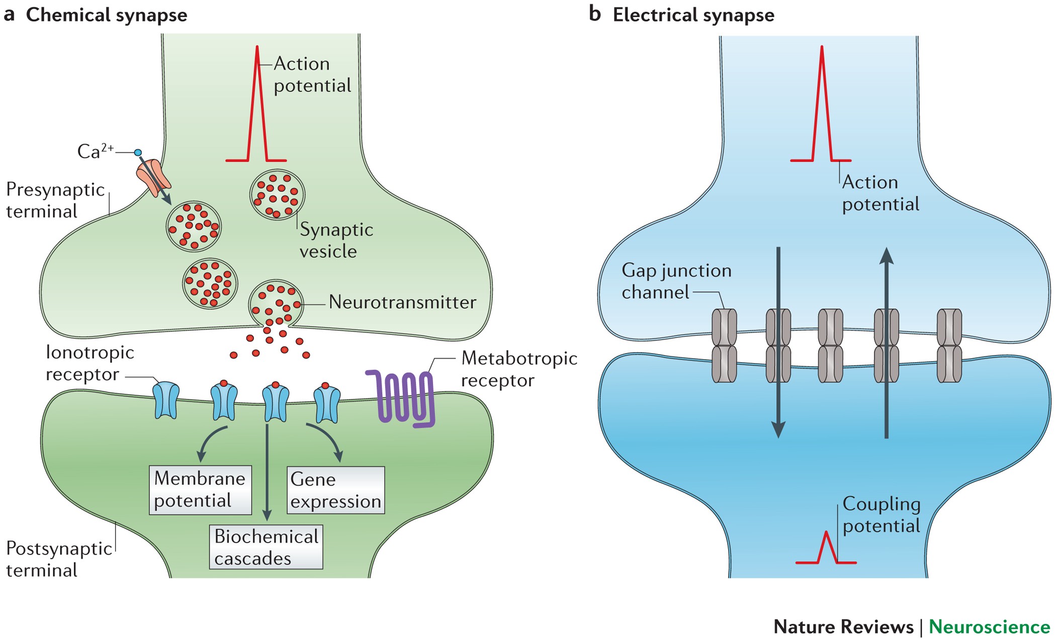 Source: Nature
Chemical synapses are the main and most adaptable way that neurons communicate in the nervous system. This process involves a series of biochemical events: when an action potential reaches the presynaptic terminal, it opens voltage-gated calcium channels, causing synaptic vesicles to release neurotransmitters into the gap between the neurons. The neurotransmitters then diffuse across this gap and bind to specific receptors on the postsynaptic neuron, changing its membrane potential. Since chemical transmission relies on diffusion and binding to receptors, it takes a little time, but it allows the signal to be strengthened, weakened, or changed as the brain needs.
Electrical synapses work differently. They connect neighboring neurons through direct physical links called gap junctions. These junctions have specialized proteins called connexons that line up between the cell membranes, allowing ions and small molecules to pass directly from one cell to another without any chemicals in between. This direct connection lets signals pass between cells almost instantly, in both directions. Electrical synapses are essential for neural circuits that need to respond quickly, such as those that control heart muscle contractions or hormone release in the brain.
"Connectivity forms the function" $\rightarrow$ Neural function comes from how synapses connect neurons, not from the neurons themselves. The strength of these connections and the paths they take determine what happens when a circuit is triggered - whether it's a reflex, a memory, or a complex thought. The way synapses are connected is what turns cell structure into brain function.
This process is still under research. Once we internalize the sheer scale of neurons in our brain, we cannot formalize how the brain handles all of it.
Synaptic Plasticity
The brain has a basic mechanism for changing itself, which is called synaptic plasticity. This process allows the connections between neurons to become stronger or weaker depending on how much they are used. Canadian psychologist Donald Hebb described this idea in 1949 with a simple rule: when neurons work together, they form stronger connections. The connections between neurons, called synapses, are not fixed. Instead, they change how well they communicate based on how often they are used. This process is the physical basis for how we learn, form memories, and adapt to new situations.
At the cellular level, this adaptation primary manifests through two opposing mechanisms:
- Long-Term Potentiation (LTP): When high-frequency stimulation persists, calcium enters through NMDA receptors. This leads to more AMPA receptors being added to the postsynaptic membrane, making the synapse more sensitive and strengthening the connection. This process forms the physical basis of long-term memories.
- Long-Term Depression (LTD): Low-frequency stimulation over a long period causes a small amount of calcium to enter, resulting in the removal of AMPA receptors from the synapse. This weakens the connection, helping to get rid of unnecessary signals and prevent neural circuits from becoming overloaded.
Together, LTP and LTD balance synaptic strength across brain structures like the hippocampus and cortex, driving cognitive processes from motor skill learning to complex reasoning.
Synaptic transmission
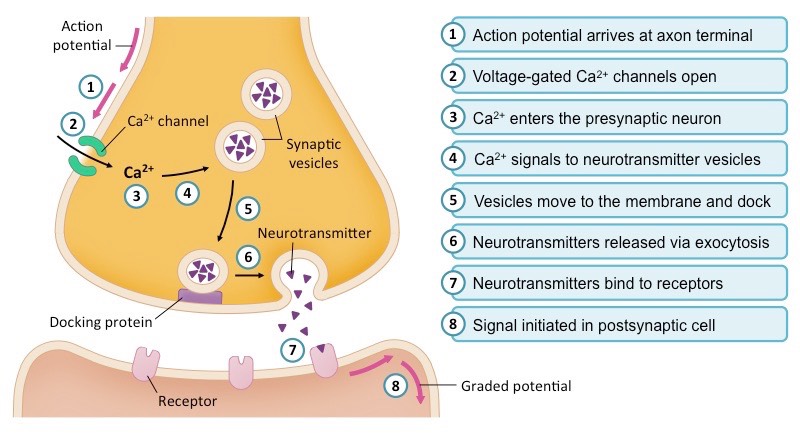 Source: BioNinja
- Arrival of the Action Potential: An electrical nerve impulse travels down the axon to the presynaptic axon terminal.
- Calcium Influx: The change in electrical voltage opens voltage-gated $\text{Ca}^{2+}$ channels in the presynaptic membrane, allowing calcium ions ($\text{Ca}^{2+}$) to rush in.
- Vesicle Docking & Exocytosis: The surge of calcium triggers synaptic vesicles filled with neurotransmitters to move to, dock with, and fuse to the presynaptic membrane, releasing their contents.
- Diffusion Across the Synaptic Cleft: The released neurotransmitters spill into the synaptic cleft and quickly diffuse across the gap.
- Receptor Binding & Signal Initiation: Neurotransmitters bind to neurotransmitter receptors on the postsynaptic membrane, located on a dendritic spine. This binding opens or closes ion channels, generating a local graded potential in the receiving neuron.
- Termination of the Signal: The remaining neurotransmitter molecules in the cleft are cleared by:
- Re-uptake pumps: Transporters return neurotransmitters to the presynaptic terminal to be recycled.
- Enzymatic degradation: Enzymes break down the neurotransmitters.
- Diffusion: Molecules drift away from the synaptic cleft.
Synaptic transmission isn't a one-size-fits-all process; where a synapse connects and how many connections exist fundamentally shape how a neuron processes information.
1. Spatial Variety
Neurons receive signals across different anatomical regions, and the location of a synapse acts like a volume control or routing mechanism for incoming information:
- Axodendritic Synapses (Axon to Dendrite / Dendritic Spine): These are the most common connections in the central nervous system. Dendrites act as the primary "receiving antennas" for signals, which often weaken as they travel from the dendrites to the soma, the cell body. These synapses allow for fine-tuned integration of excitatory or inhibitory inputs.
- Axosomatic Synapses (Axon to Cell Body / Soma): Synapses on the soma have a strong influence on whether the postsynaptic neuron fires an action potential. The soma is close to the axon hillock, where action potentials originate, so synapses here can effectively control the neuron's firing. These inputs are often inhibitory and can prevent the neuron from firing.
- Axoaxonic Synapses (Axon to Axon): These connections affect how neurons communicate with each other. Instead of changing the voltage of the postsynaptic cell, they regulate how much neurotransmitter the target neuron can release. This can either inhibit or facilitate the release of neurotransmitters.
2. Synaptic Strength & Multiple Contact Points
A single input from one synapse is rarely enough to force a receiving neuron to fire an action potential. Neurons rely on the summation of multiple contact points and variable synaptic strength to compute decisions:
- Multisite Connections: A single presynaptic axon often forms several physical contact points with the same postsynaptic cell. This increases the reliability of signal transmission, ensuring that an action potential in the sending cell releases enough total neurotransmitter to produce a response.
- Spatial and Temporal Summation:
- Spatial Summation: When multiple separate synapses fire simultaneously at different locations on the same receiving cell, their individual electrical currents combine at the axon hillock.
- Temporal Summation: When a single synapse fires repeatedly in rapid succession, the electrical charges build on top of one another before the cell can return to rest.
- Modulating Synaptic Strength: Synapses are not all equal. The strength of a synapse depends on how much neurotransmitter is released, how many receptors are present on the receiving side, and the structural features, such as the shape of dendritic spines, that affect electrical current. Strong synapses have a major influence during summation, while weaker synapses provide subtle background inputs.
Together, the placement and multiplicity of synapses allow a neuron to function as a computer, weighing hundreds of varied inputs to produce a single, unified output.
Neurotransmitters
Neurotransmitters are chemical messengers that help neurons communicate with each other at synapses. They are made in the neuron and stored in small vesicles. When calcium ions flow in, these chemicals are released into the space between the neurons. They then cross this gap and attach to receptors on the neighboring neuron's membrane. This attachment changes the electrical properties of the receiving neuron, influencing a wide range of functions, from simple reflexes to complex thought processes.
- Glutamate: The principal excitatory neurotransmitter in the central nervous system. It depolarizes postsynaptic cells to promote action potentials and serves as the primary driver of synaptic plasticity, learning, and memory.
- GABA ($\gamma$-Aminobutyric Acid): The main inhibitory neurotransmitter in the brain. It hyperpolarizes postsynaptic membranes to reduce neuronal excitability, preventing overstimulation and maintaining neural stability.
- Acetylcholine (ACh): Operates at neuromuscular junctions to trigger muscle contraction and acts as a key neuromodulator in the brain to regulate arousal, attention, and memory encoding.
- Dopamine: A vital neuromodulator that governs reward-seeking behavior, motivation, motor control, and executive decision-making circuits.
- Serotonin (5-HT): Regulates mood, emotional processing, sleep-wake cycles, appetite, and gut-brain signaling.
- Norepinephrine (Noradrenaline): Mobilizes the brain and body for action by increasing alertness, focus, and vigilance during stress or environmental changes.
- Epinephrine (Adrenaline): Primarily drives peripheral "fight-or-flight" sympathetic responses, elevating heart rate and blood flow during acute stress.
- Endorphins: Neuropeptides that function as natural analgesics, binding to opioid receptors to block pain signaling and induce euphoria.
Key Types of Postsynaptic Receptors
The action of a neurotransmitter is ultimately determined not by the chemical itself, but by the specific receptor it binds to on the postsynaptic side:
- Ionotropic Receptors (Ligand-Gated Ion Channels): Direct, fast-acting receptors where the binding site and ion channel are part of the same protein complex. Binding immediately opens an ion channel, generating fast electrical responses lasting only milliseconds (e.g., AMPA, NMDA, and $\text{GABA}_\text{A}$ receptors).
- Metabotropic Receptors (G-Protein Coupled Receptors / GPCRs): Indirect, slower-acting receptors that operate through intracellular G-proteins and second-messenger cascades. Instead of immediately opening a channel, binding triggers long-lasting cellular changes, altering overall excitability and gene expression over hundreds of milliseconds to minutes (e.g., Muscarinic ACh, $\text{GABA}_\text{B}$, and Dopamine receptors).
Action Potentials
 Source: Zoë Huggett
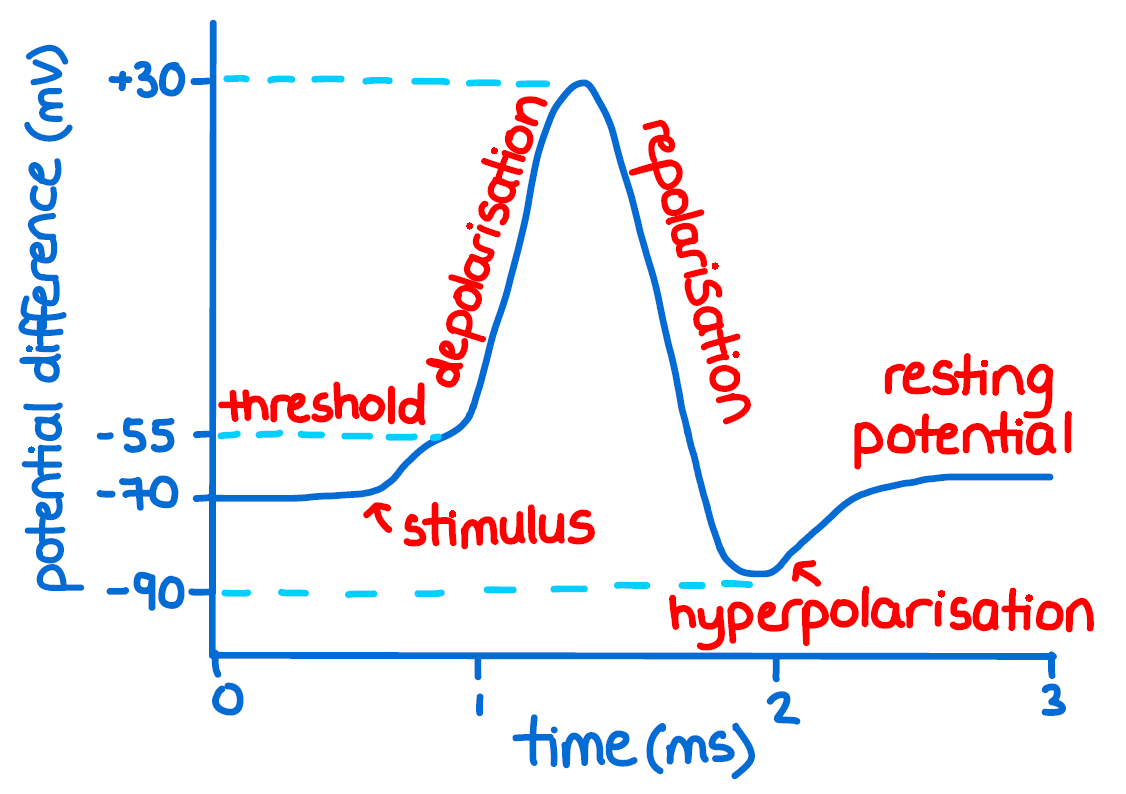
An action potential, also known as a spike or nerve impulse, is a rapid, temporary electrical discharge that travels along a neuron's membrane. These spikes are the main signals used by the nervous system to process sensory information, control movements, and perform complex calculations involved in learning and memory.
The state of a neuron's membrane is defined by its membrane potential, which is the voltage difference between the inside and outside of the cell. This is represented by the equation:
$$V_i - V_0 = V_{\text{mem}} = \Delta V = V$$
When a neuron is at rest, it has a negative charge compared to its surroundings, usually between $-60\text{ mV}$ and $-70\text{ mV}$. The neuron maintains this resting state through ion pumps and selective channel permeability, which mainly control the balance of sodium, potassium, and chloride ions.
Excitation vs. Inhibition
Incoming synaptic inputs change the membrane potential, moving the cell closer to or further from producing a spike. This happens in two ways:
- Excitation increases the positivity of the membrane potential. For example, it might change from $-70\text{ mV}$ to $-55\text{ mV}$. If the voltage reaches a certain threshold, the cell generates an action potential.
- Inhibition increases the negativity of the membrane potential. For example, it might change from $-70\text{ mV}$ to $-90\text{ mV}$. This stabilizes the cell and prevents it from producing a spike.
Stages of Action Potential An action potential occurs as an "all-or-none" event, driven by voltage-gated ion channels, and consists of seven distinct phases:
- Resting State: The cell remains at its resting potential, around -60 to -70 millivolts. At this time, voltage-gated sodium and potassium channels are closed.
- Initial Depolarization: When the cell receives excitatory inputs, the voltage shifts upward. This happens because a small number of voltage-gated sodium channels open, allowing sodium ions to enter the cell.
- Threshold: If the voltage reaches the threshold voltage, around -55 millivolts, many more voltage-gated sodium channels open across the membrane. This creates a positive feedback loop.
- Rising Phase (Upstroke): As a result of the opening of these sodium channels, a large number of sodium ions rush into the cell. This causes the membrane voltage to surge upward rapidly, peaking at around +30 to +40 millivolts.
- Falling Phase (Repolarization): The voltage-gated sodium channels then quickly inactivate, and voltage-gated potassium channels open. Potassium ions rush out of the cell, driving the voltage back down toward negative values.
- Undershoot (Afterhyperpolarization): Because the potassium channels close relatively slowly, potassium ions continue to leave the cell for a brief time. This causes the voltage to drop lower than the resting baseline, more negative than -70 millivolts.
- Refractory Period: During this brief period, the neuron is unable to fire another action potential easily. The absolute refractory period, during which sodium channels are inactivated, prevents the spike from traveling backward. This ensures that the signal travels in one direction along the axon. The relative refractory period requires a much stronger excitatory signal to trigger a subsequent spike.