Neuron Structure
Table of Contents
This is a series of blogs on Computational Neuroscience. To get a heads up, let's define the subject area:
Computational neuroscience is a field that combines different disciplines to study the brain using mathematical models, computer simulations, and theoretical analysis. This approach helps us understand how the brain develops, processes information, and controls behavior, connecting biological data with the computational principles that govern neural systems.
In simple terms, it's the study of $\text{neuroscience} + \text{modelling brain activity}$. To better understand this area, I'm going to build the spectrum piece by piece. Today, it's Neuron.
Neuron are the foundational communication blocks of the nervous system. When we think, feel, or move, neurons send electrical impulses and chemical signals to each other. The brain then acts as a command center, organizing the constant communication between these cells. As billions of neurons connect and fire together, the brain processes information, stores memories, and creates everything that makes us human.
Brain
For scientific study of Brain, it's structure is mapped from 3D to 2D. They are;
- Horizontal or axial plane (Also called as transverse section)
- Coronal or frontal plane
- Sagittal plane
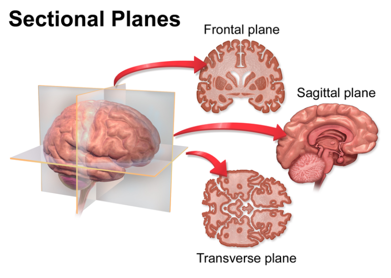 Image Courtesy: Rotel Project
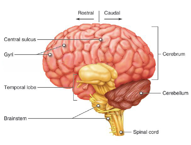 Image Courtesy: Lumen Learning
The brain's cortex has a layered structure. Each of its six layers is different and contains a specific mix of neuron types, with distinct shapes and connections. This organization determines what each layer does. Layer 4 is particularly important as it receives most of the sensory information and other complex inputs that come from deeper parts of the brain, especially the thalamus. It acts as the main entry point for the cerebral cortex.
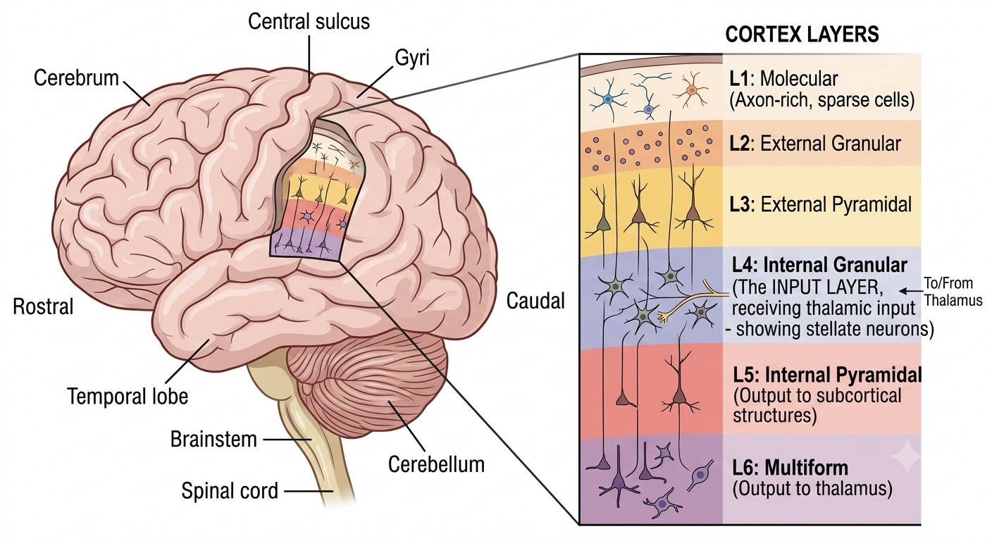 Layered structure is generated using AI
Neurons Structure
To study Neurons scientists have to use staining techniques to visualize the structure and patterns of neurons in the brain. This work began with Camillo Golgi's method, which uses silver nitrate to stain a small number of neurons completely, showing their different parts like axons and dendrites. Santiago Ramón y Cajal used this technique to map the nervous system. Now, neuroscientists use fluorescent stains and genetic labeling, such as "Brainbow", to create detailed maps of neural circuits. These maps allow researchers to follow specific connections, study the structure of neurons, and understand how these cells process information.
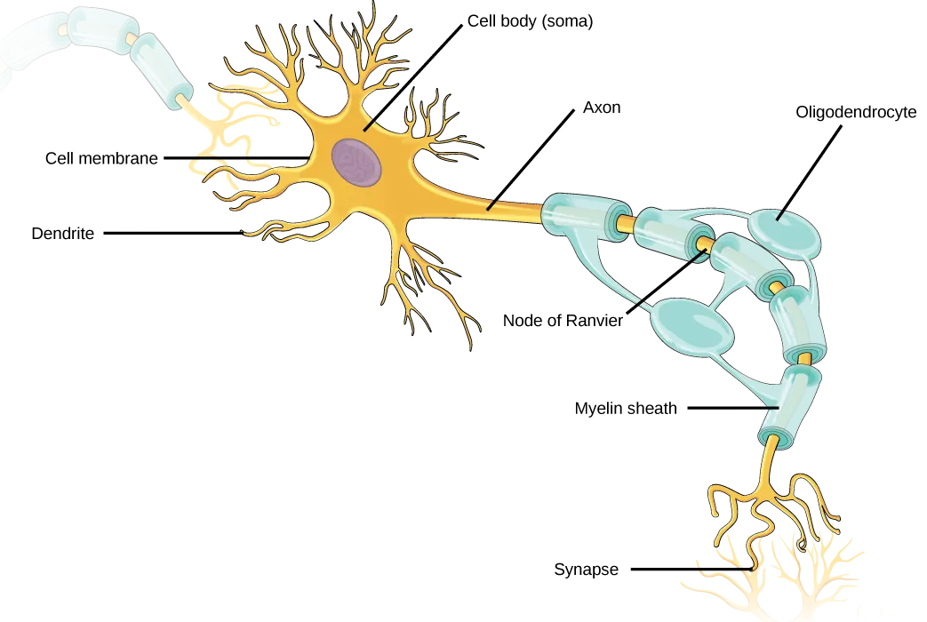 Courtesy: Openstax
A neuron has three main parts: the cell body, dendrites, and an axon. The cell body, also called the soma, contains the nucleus and controls the cell's metabolic functions. Dendrites branch out from the cell body and receive chemical signals from nearby neurons. The signals are then integrated and sent down the axon, a long extension that carries electrical impulses away from the cell body. The axon is covered with a myelin sheath, made up of light blue segments wrapped around a gold core, which helps speed up signal transmission. This sheath is produced by supporting glial cells and allows the signal to travel quickly down the axon until it reaches the end, where it is passed on to other neurons.
A standard neuron has a clear input and output side. The dendrites receive incoming chemical messages, while the axon transmits the signal away. The axon hillock, a region between the cell body and the axon, is where the neuron integrates incoming electrical signals and decides whether to fire a nerve impulse. This region acts as the starter for the action potential. Dendrites often have thousands of small protrusions called dendritic spines, which increase the surface area for connections and act as individual sites for specific inputs. However, not all neurons have this structure - some lack dendrites or axons and use different designs to sense their environment and communicate.
Foundational Work
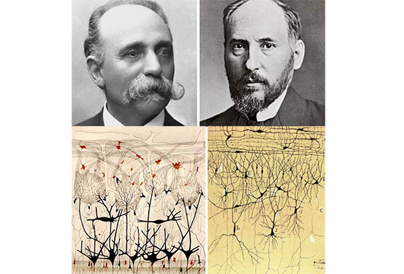 Image Courtesy: Eldiariodesalud
In the late 19th century, the scientific community was divided over how the nervous system processed and transmitted information. Golgi proposed the Reticular Theory, which held that the nervous system was a continuous, interconnected web, similar to the circulatory system. He believed that the cells in this network were connected and that axons joined together to form a network where electrical signals could flow in all directions.
Cajal had a contradictory view, he used Golgi's staining technique with great precision and came to the opposite conclusion, saying that neurons are separate, individual entities. Cajal argued that instead of being physically connected, neurons are close together and communicate across tiny gaps, which were later named synapses. This debate went on for decades until the 1950s, when the electron microscope was able to show the physical gap of the synaptic cleft for the first time, proving Cajal was right.
Cajal's work laid the foundation for modern neuroscience with the Neuron Doctrine, which provided a framework for understanding brain function. By showing that neurons are individual units, scientists could categorize them by shape and function and map specific neural pathways. Cajal also discovered the Law of Dynamic Polarization, which states that signaling in a neuron is strictly one-way: information enters through the dendrites and cell body, travels down the axon, and is transmitted to the next neuron. This orderly process could not be explained by Golgi's idea of a continuous network. The idea that the brain is made up of individual, interacting neurons is now a cornerstone of fields like computational neuroscience, where the brain is treated as a network of separate nodes rather than a single, continuous mass.
The rivalry between the two scientists came to a head in 1906, when they were jointly awarded the Nobel Prize in Physiology or Medicine for their work on the nervous system. The award highlighted an interesting twist: Cajal had used a chemical method developed by Golgi to challenge Golgi's main theory. In 1873, Golgi had discovered a silver nitrate staining method known as the "black reaction," which stained some neurons completely, making them stand out against a clear background. Cajal improved on this technique, using it to map the paths of axons and dendrites with great accuracy. The tension between the two men was still evident at the Nobel ceremony, where Golgi used his acceptance speech to strongly defend his Reticular Theory. The next day, Cajal gave a lecture that carefully refuted Golgi's arguments, using detailed, hand-drawn diagrams to make his points. Although Golgi had developed the crucial staining method, it was Cajal's insightful work that laid the groundwork for modern neurobiology.